The IUCr Newsletter is published quarterly and distributed free of charge to 587 libraries and 15,000 crystallographers and other interested individu als in 39 countries. Feature articles, meeting announcements and reports, information on research or other items of potential interest to crystallogra phers should be submitted to the editor at any time. Submission of text by electronic mail and graphic slides or photographs by express mail re quested. Items will be selected for publication on the basis of suitability, content, style, timeliness, and appeal. The editor reserves the right to edit. The Newsletter is published by the International Union of Crystallography, 2 Abbey Square, Chester CH1 2HU, UK. Cost of distribution in Australia, Czech Republic, France, India, Italy, Japan, Poland, New Zealand, South Africa, Switzerland, and The Netherlands is borne by crystallographic associations or institutes or by individual crystallographers in these countries.
Address changes or corrections and request to be added to the mailing list should be addressed to the edito rial office.
Send Contributions to:
W.L. DuaxTel.: 716-856-9600 · FAX: 716-852-4846
e-mail: patti@hwi.buffalo.edu
Matters pertaining to advertisements should be addressed to W.L. Duax or P. Coley at the above address.
In Japan, contact:
Prof. Yohio MitsuiDept. of Bio Engineering
Nagaoka U. of Technology
Nogaoka, Niigata 940-21, Japan
FAX: 81-258-46-8163.
Everywhere else, contact W.L. Duax or P. Coley at the Buffalo office.
William L. Duax
Editor
Patti Coley
Copy Production Manager
Jane Griffin
Assistant Editor
Linda Katz
Newsletter Design & Production
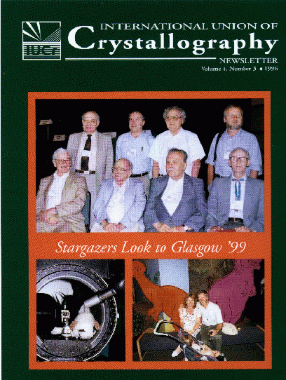
On the Cover: Astrological Signs predict major scientific happening in Glasgow in the summer of 1999. The blessed event is being planned by program chair Judith Howard and Local Chair Chris Gilmore. Shown in the photo on the lower right-hand corner of the cover. There will be plenty of mutton on hand. More than eight wise persons are expected to follow the rising star of crystallography to Glasgow. The eight Nobel Laureates shown on the cover (Standing: J. Karle, H. Deisenhofer, H. Hauptman, H. Michel, seated: J. Kendrew, C. Shull, B. Brockhouse, W. Lipscomb) attended the XVII Congress in Seattle.
Contributors: A. Albinati, A. Authier, T. Baker, D. Brown, C. Bugg, S.K. Burley, K. Cowtan, H. Einspahr, P. Fitzgerald, H. Flack, R. Fletterick, S. Fortier, R. Fourme, C. Gilmore, E. Goldsmith, A. Golobic, M. Grutter, M. Hart, J. Helliwell, W. Hol, S. Hovmoller, L. Jensen, L. Johnson, J. Kelly, J. Lipkowski, J.M. Newsam, H. Novoa de Armas, M. Sakata, J. Simpson, J. Smith, R. Sweet, M. Van Hove, J.-L. Verger-Gaugry, D.J. Watkin.
The IUCr Newsletter (ISSN 1067-0696; coden IUCNEB) Volume 4, Number 3. Published quarterly (4x) by the American Institute of Physics for the International Union of Crystallography. Members receive the IUCr Newsletter by virtue of their membership in the IUCr. Periodical postage rates paid at Woodbury, NY and additional mailing offices. POSTMASTER: Please send changes of address to IUCr Secretariat, 2 Abbey Square, Chester, CH1 2HU, England.
Table of Contents
Letter From The President
Letter From The Editor
Letters to the Editor
IUCr Activities
IUCr Commissions (19961999)
CIFNEWS
Maximum Entropy Method Project
Your Opinion Please
IUCr Congress in Glasgow
IUCr Congress Report
Meeting Reports
X-Ray and Neutron Dynamical Diffraction
Theory and Applications
Powder Diffraction Cuba
Going MAD in Grenoble
Regional Affiliate News
Asian Crystallographic Association News
ACA 1997 Meetiing
Future Meetings
Way Down Under
BCA Spring Meeting
Erice 97, May 22June 2
Organic Crystal Chemistry
Aperiodic '97
Meeting Calendar
Hope you enjoy our first attemp at online distribution. Look for photos in the Spring online version!
Letter From The President
Reflections on Seattle
It was exciting scientifically and it was one of those truly international occasions through which we meet up with old friends and make many new ones.
Much credit goes to the organizers who devised the program and provided a venue with space for people to mingle, or view the posters and displays at leisure, when not attending lectures.
For me the Congress also brought about my election as President of the Union for the next three years. This was a very great honor, not just for me but for New Zealand and for the many smaller countries that are a vital part of the Union. It is also somewhat daunting, but I am very encouraged by all the good wishes and expressions of support I have received from friends around the world. At home my research group even produced a special Tshirt!
In this first letter I thought it appropriate to make some comments on the nature of our triennial Congress. It has become very large (more than 2500 participants in Seattle). It is also very broad, with so many different specialities represented. This inevitably leads to suggestions that it is too large or that particular areas are not given sufficient recognition in the program.
My own feeling is that our breadth is a great strength, and that it is very important that we have this one special occasion on which we all meet together. This was brought home to me particularly in some of the macromolecular sessions. The Seattle Congress brought a breathtaking array of new biological structures with profound implications for biology and medicine. Some of these were recently highlighted in Science (August 30, p. 1174 "Crystallographers pinpoint what goes where"). What struck me, however, was that the sessions on methods generated enormous interest, emphasizing that it is the crystallographic method that underlies all these achieve ments. In macromolecular crystallography, just as in other areas, methods constantly evolvethe growing importance of synchrotron radiation and of electron diffraction are but two examples.
Sir John Kendrew, in one of the Nobel Lectures, reminded us that it is the problem to be solved that determines what methods should be used. Our triennial Congress is then essential for the crossfertilization that should result when so many crystallographers from different areas, with different approaches, are brought together. In the end, too, I hope that we all draw tremendous pleasure from some of the spectacular achievements that are presented. They vividly demonstrate the vitality and power of crystallography.
Members of the IUCr Executive Committee
19961999
R. Chidambaram Vice-President
S. Larsen Gen. Sec. & Treasurer
P. Coppens Past-President
L.A. Aslanov J.C.A. Boeyens
H. Fuess M. Hart
H. Schenk M. Tanaka
XVII Congress Report
This issue of the IUCr Newsletter contains highlights of some of the reports on scientific sessions at the
XVII Congress and General Assembly of the IUCr held in Seattle Aug. 917.
The full reports on 13 keynote addresses and 62 microsymposia written by
the session chairs have been placed on the meeting website: http://www.hwi.buffalo.
edu/aca/. Additional reports are being added as they are received. Because
of the page limits, not all of the reports can be printed in the Newsletter
and those that are have to be edited.
This issue contains reports on keynote addresses and microsymposia related to instrumentation and experimental techniques, methods of structural determination, computers and modeling, and macromolecular crystallography. Other congress topics will be covered in the next two issues of the newsletter.
The record congress attendance of 2713 crystallographers from 51 countries, included 252 accompanying members and 150 exhibitors. A detailed statistical report concerning the geographical distribution of abstract submissions and meeting attendance will appear in a future issue. A summary report on the opinions concerning the formats and content of future IUCr congresses expressed by 485 congress participants who completed questionnaires distributed on the morning of the last full day of the congress will also appear in a future issue.
Industrial Support
A record number of exhibits contributed to the scientific and financial success of the Seattle meeting. Many of the same exhibitors advertise regularly in this newsletter. It is only through the generosity of the International Union of Crystallography and with the support of the advertisers that the newsletter can be produced and sent to you at no cost. If each reader were to send a letter, FAX, or e-mail message to just one advertizer in the next month expressing their appreciation to them for supporting crystallography and the newsletter it could make a significant difference to the long term survival of the newsletter.
IUCr Activities
The IUCr Newsletter is an ideal vehicle for alerting the International Community to the activities of the Commissions of the IUCr and its regional affiliates. A report from the Commission on Charge, Spin, and Momentum Density appears on page 6 and a report from the Asian Crystallographic Association, a regional affiliate of the IUCr, appears on page 20.
Meeting Reports
Page limits have created a backlog of scientific meeting reports. Some national and regional newsletters ( BCA News, ACA News) do such an excellent job of providing in depth coverage of local meetings that only highlights can be incorporated into the IUCr Newsletter. An effort is made to include meeting reports that sample the entire range of crystallographic interests and geographical areas.
Obituaries
We are saddened by the news of the deaths of many prominent crystallographers including Boris Konstantinovish Vainshtein (Russia), Harry Cailisle (UK), Sir Gordon Cox (UK), Mel Mueller (USA), David Schomaker (USA), and Stan Simonsen (USA). Brief accounts of their lives will appear in future issues.
I thought the organizers of the IUCr Congress in Seattle might like to know that after my abstract appeared on the Web, together with my e-mail address, I received several e-mail messages from people who had spotted the abstract. In more than one case, these were from people not attending the congress who otherwise would not have been aware of our results. In one case, this has led to a future collaborationas a direct result of the abstracts being available on the Web pages. Thanks to all involved.
I agree with your recent comments on the work by Tegze et al. and Gog et al. on X-ray holography at atomic resolution. Indeed, readers of the many reviews of the recent work on X-ray holography could be forgiven for believing that Gabor's 1948 Nobel-prize winning proposal was for X-ray holographic imaging of atoms, and that this has at last been achieved. This impression is grossly misleading because Gabor did not propose X-ray holography, and the new experimental results are diffraction patterns and not holo grams. Gabor proposed electron holography, not X-ray holography, for the purpose of elliminating electron microscope aberrations. Since the first implementation of Gabor's proposal by Haine and Mulvey in 1952 this field has made steady progress, demonstrating both spectacular images of superconducting vortices in motion, and atomic resolution images of defects in crystals.
For the more limited case of diffraction experiments involving periodic structures which recent work describes, the use of the scattering from particular types of atoms to form reference beams has its origins in Bragg's two-wavelength X-ray microscope of 1942, which formed optical reconstructions of diopside from X-ray diffraction patterns, using heavy atoms to solve the phase problem. For the more challenging case of an isolated, non-periodic atomic structure, X-ray holography probably started with the remarkable optical reconstruction of the X-ray image, (showing four Fresnel fringes) of a needle by El-Sum and Kirkpatrick in 1952. Again, continuous progress in this field of true X-ray holography has been made since then using synchrotrons by researchers such as Jacobsen, Kirz, and Howells in this country.
The "new" XFH scattering distribution is not a hologram because the reconstructed wavefield is not conjugate to the objectit is not an image as Gabor clearly intended. Because the method assumes crystallographically equivalent emission sites, it is much more similar to the conventional Kossel diffraction patterns analyzed by von Laue in 1935. (Von Laue gave the theory for both XFH and MEXH, which are related by reciprocity, in 1935.) The MEXH technique continues a tradition of work begun by Knowles in 1956, who studied the effect of neutron standing waves in crystals on X-ray emission, and continued by Batterman, Bedzyk, Duncumb, and many others, including the authors, who made the connection with structural analysis [ Acta Cryst. 17, p.33 (1964); J. Micros. 130, p.147 (1984)].
Both the recent synchrotron experiments are remarkable experimental achievements, for which the tunability and intensity of the sychrotron provides exciting new possibilities. But computer modelling of diffraction data must be distinguished from Gabor's holog raphy. By creating a spurious impression of novelty and confusing the distinct topics of diffraction and holography, readers may be misled, and deprived of the rich history of these subjects.
In your editorial on holography with X-rays, you state that the homometric solution belongs in the same category as the Loch Ness monster and the Abominable Snowman. If you write to any major museum of natural history, I am sure that a request for even the
thinnest slice of the mentioned species will bring you a negative reply. So why not ask for a sample of the mineral tapiolite FeTa 2O6 instead? The structure of this mineral is simply a three-fold superstructure of the rutile type and tapiolites with partial cation disorder appear to exhibit homometry [Acta Cryst, (1995), A51, 514519]. Our results on tapiolite can probably be extended to other three-fold superstructures caused by cation ordering, of which there are many known examples among minerals and ceramic materials. Even if homometric structures turn out to be so rare that X-ray crystallographers need not really wor ry, I think that little is gained by pretending they do not exist at all.
My remarks with respect to homometric solutions were made in reference to the undue emphasis placed upon the phenomenon by early opponents of direct methods who believed the work of Hauptman and Karle was the heretical ravings of upstart mathematicians and physicists with little understanding of chemistry and the laws of crystallography. The fearful specter raised was that direct methods would lead to an incorrect solution, that was chemical nonsense but mathematically indistinguishable from a correct one and that correct solutions would be missed. As I understand it your "homometric structures" are partially disordered atomic arrangements that satisfy the same diffraction pattern. I believe our understanding of what "homometric" means is colored by our experience.
I recently had a paper rejected by the Journal of the American Chemical Society (JACS) on the grounds that the policy of the journal is to no longer accept papers based on interpretation or reinterpretation of existing data. The work in question was based on data from the CSD and otherwise got a favorable review. Therefore I wrote the letter reprinted below. Due to C & E News policy of publishing only letters related to stories that appear in C & E News itself, my letter will not be published, I thought the readers of the IUCr Newsletter should be alerted to this policy.
Letter to the Editor C&E News:
Many of your readers, potential authors of papers submitted to the Journal of the American Chemical Society, will be interested to know that it is now JACS policy not to publish papers that primarily take existing published literature data and use it without significant new data, experimental or theoretical contributions. This ruling applies not only to results based on so-called quantitative structure -activity relations (QSAR), chemometric treatment of data sets, etc., but also to those based on systematic studies of published crystal structures collected from databases such as the Cambridge Structural Database (CD) or the Brookhaven Protein data bank (PDB). A recent referee's report from a member of the JACS editorial board states that "JACS should not be the venue for (re)interpretation of existing data."
This means that papers dealing with the systematics of intermolecular interactions as expressed in crystal packing patterns are to be excluded. Of the thousands of crystal structures that are determined annually, only a tiny fraction receive any discussion whatsoever in the published literature, and in these it is usually the molecule itself that is the center of discussion; the packing is hardly ever men tioned. Besides, what can be learned from a study of the packing in one crystal structure? One has to look at many to identify the recurring patterns, the exceptions from these, and the reasons for these exceptions.
In the past, the (re)interpretation of existing data has played a vital role in the development of chemistry. Are we now to limit what is important only to the latest (truly novel) experimental results? One should keep in mind that according to the new ruling, many of Pauling's important papers might not have qualified for JACS. Mendeleev's paper on the periodic system would almost certainly have been rejected.
IUCr Commissions (19961999)
G.C. Chapuis (CH) (Chair)
M. Farkas-Jahnke (HU)
J.M. Dubois (FR)
D. Pandey (IN)
J.M. Pérez-Mato (ES)
M.L. Senechal (US)
W. Steurer (CH)
A. Yamamoto (JP)
S. Van Smaalen (DE)
BiologicalMacromolecules
W.H.E. Saenger (DE) (Chair)
M. Bolgnesi (IT)
W.-r. Chang (CN)
J.K. Dattagupta (IN)
A. Liljas (SE)
Y. Mitsui (JP)
J. Sevcik (Slovakia)
J.L. Smith (US)
I. Wilson (US)
Charge, Spin,and Momentum Densities
K. Schwarz (AT) (Chair)
A. Bansil (US)
F. Itoh (JP)
G.H. Lander (DE)
C. Lecomte (FR)
F. Sacchetti (IT)
M. Sakata (JP)
M.A. Spackman (AU)
V. Tsirelson (RU)
Y. Wang (Taipei)
Crystal Growth and Characterization of Materials
H. Klapper (DE) (Chair)
P. Bennema (NL)
P.M. Dryburgh (UK)
F. Licci (IT)
N.-B. Ming (CN)
T. Nishinaga (JP)
F. Rosenberger (US)
P. Rudolph (DE)
I. Smolsky (RU)
Crystallographic Computing
P. Bourne (US) (Chair)
G.L. Cascarano (IT)
T. Higashi (JP)
M. Kjeldgaard (DK)
T. Koritsanszky (DE)
M. Ramanadham (IN)
J. Simpson (NZ)
A.L. Spek (NL)
D. Watkin (UK)
Crystallographic Nomenclature
S.C. Abrahams (US) (Chair)
Ex officio members:
Editor-in-Chief of Acta Cryst.
Section Editors of Acta Cryst.
Editor of J. of Applied Crystallography
Main Editors of J. of Synchrotron Radiation
Editors of Int'l Tables for Crystallography
Chair of IUCr/OUP Book Series Comm.
Chair of Crystallographic Teaching
Crystallographic Teaching
C.M. Gramaccioli (IT) (Chair)
L.A. Aslanov (RU)
C.P. Brock (US)
I.D. Brown (CA)
P.N. Kasai (JP)
E. Makovicky (DK)
Y.P. Mascarenhas (BR)
A. Oskarsson (SE)
P. Phavanantha (TH)
W.T. Robinson (NZ)
Electron Diffraction
J.W. Steeds (UK) (Chair)
D.L. Dorset (US)
A. Ichimiya (JP)
F.-h. Li (CN)
J.C.H. Spence (US)
M. Tanaka (JP)
M.A. van Hove (US)
D. Van Dyck (BE)
High Pressure
R.J. Nelmes (UK) (Chair)
Y. Fujii (JP)
I.N. Goncharenko (RU)
D. Hausermann (FR)
R.J. Hemley (US)
A. Katrusiak (PL)
W.F. Kuhs (DE)
J.B. Parise (US)
O. Shimomura (JP)
Neutron Scattering
J.W. White (AU) (Chair)
A.M. Balagurov (RU)
C.J. Carlile (UK)
J.B. Forsyth (UK)
Y. Fujii (JP)
P.S. Goyal (IN)
G. Heger (DE)
J.D. Jorgensen (US)
J.S. Pedersen (DK)
E. Prince (US)
Powder Diffraction
R.J. Cernik (UK) (Chair)
R. Delhez (NL)
L.B. McCusker (CH)
J. Pannetier (FR)
P. Scardi (IT)
S.P. Sen Gupta (IN)
D.K. Smith (US)
I.G.R. Tellgren (SE)
H. Toraya (JP)
R. Von Dreele (US)
Small Angle Scattering
J.D. Barnes (US) (Chair)
A. Craievich (BR)
E. Kaler (US)
G. Kostorz (CH)
K. Osamura (JP)
J. Penfold (UK)
T.M. Sabine (AU)
D. Svergun (RU)
Structural Chemistry
C. Krüger (DE) (Chair)
V.K. Belsky (RU)
G. Desiraju (IN)
J. Flippen-Anderson (US)
G. Gilli (IT)
K. Hagen (NO)
B. Kojic-Prodic (HR)
B. Krebs (DE)
Y. Ohashi (JP)
G. Punte (AR)
Synchrotron Radiation
Y. Amemiya (JP) (Chair)
R. Feidenhans'l (DK)
A. Fontaine (FR)
K.K. Kannan (IN)
A.H. Kvick (FR)
D.P. Siddons (US)
K.D. Watenpaugh (US)
S.W. Wilkins (AU)
A. Yonath (IL)
XAFS
E.A. Stern (US) (Chair)
K. Baberschke (DE)
A. Fontaine (FR)
K. Garg (IN)
B. Hedman (US)
S. Mobilio (IT)
T. Murata (JP)
J. Penner-Hahn (US)
Complete addresses can be found in the IUCr World Directory on line at http://www.iucr.ac.uk
It is nearly five years since Acta Crystallographica C started accepting papers electronically as Crystallographic Information Files (cifs). During that time cif has become a recognized standard adopted by all the major structure-solving packages. The recent 10% per year increase in the submission rate of papers to Acta Cryst. C shows that authors find the format a convenient way to submit their crystal structure for publication. Since the programs checkcif and printcif are publicly available on e-mail, it is easy for authors to check their cifs and to see what their paper will look like in print before the paper is submitted. They just e-mail their cif to checkcif@iucr.ac.uk or printcif@iucr.ac.uk and the results are returned within a few minutes.
However, crystallographers who think that cif is nothing more than a convenient way to submit structure reports to Acta Cryst. do not realize that this is just the tip of the iceberg. Those who skipped the excursion at the IUCr Congress in Seattle to attend the cif workshop were treated to a review of the large amount of work that has been going on behind the scenes. Soon to be published are a much needed addition to the original (core) dictionary and a dictionary for powder diffraction that will allow Rietveld refinements to be submitted electronically to Acta Cryst. C and powder patterns to be submitted to the Int'l Center for Diffraction Data. A major dictio nary for macromolecular crystallography is near completion and will do for Acta Cryst. D and the Protein Data Bank what the core dictionary has done for Acta Cryst. C and the Cambridge Data Base. In a less advanced state are cif dictionaries for symmetry and modulated structures, and discussions are underway to create standards for area detector images and graphics. Complementing all these new dictionaries is an array of new software to edit and manipulate cifs. When all these projects come to fruition, cif will be not just a format for reporting the crystal structures of small molecules, but a standard that underpins a sophisticated crystallographic data han dling system designed to allow crystallographers to exploit the full potential of modern computers on the internet. To find the latest information about cif visit the cif home page (http://www.iucr.ac.uk/cif/home.html) where you will find documentation on cif, links to existing dictionaries, and pointers to work in progress on different aspects of cif.
Watch cifnews in future issues of the IUCr Newsletter for further information on these projects and how they will affect you.
Chair of Comcifs
The goals of a project on the Maximum Entropy Method (MEM) and a project on multipole refinements of the IUCr Commission on Charge, Spin, and Momentum Densities (CSMD) is to test the value of MEM for the study of CSMD. Objectives of the project include the following:
1. Survey the state of the discipline. Establish a system to gather information on recent results and computer programs.
2. Analyze the results. Is the same result obtained when a set of data is analyzed by different groups, using different programs and/or applying the programs in different ways? Establish criteria for weighing schemes, appropriate initial densities, and suitable experimental conditions for MEM analysis.
3. Identify the most efficient algorithms for MEM calculation and determine program limitations.
4. Analyze MEM densities in terms of models thus allowing a quantitative description of the underlying density. Characterize densities by topological properties.
5. Determine how to estimate the accuracy of the MEM charge densities and avoid pitfalls and artifacts.
In order to guide the use of MEM, it is desirable to have a better understanding of its theoretical background. Although philosophi cal discussions of MEM are outside the scope of the project, it is hoped that the project will lead to better use of MEM in other applications as well.
Those interested in participating should contact a steering committee member:
M. Sakata ( Chair), a40366a@nucc.
cc.nagoya-u.ac.jp; M. Takata (Sec.), a41024a@nucc.cc.nagoya-u.ac.jp; Members: F. K. Larsen (Co-chair), R. J. Papoular, W. Jauch; Observer: D. Feil; Advisors: G. Bricogne, D. M. Collins. The MEM calculation program, MEED is available
by an anonymous ftp: (ftp://hod.nuap.nagoya-u.ac.jp/pub/ftp.html). There
are no restrictions on methods of MEM data analysis or interpretation, and
publica tion by participants will not be restricted.
Your Opinion Please
As the newly elected Editor-in-Chief of Acta Crystallographia , I welcome comments from members of the crystallog raphic community on the Journal. There are many trends and challenges to face both in terms of publishing, such as the Internet and other electronic media, as well as scientifically, such as the increase in the numbers of "chemical" and "biological" structures being deter mined. Within the IUCr there is close cooperation already between the Commission on Journals and the IUCr's Electronic Publishing Committee where issues addressed include electronic submission, production and delivery of papers, and charging mechanisms. As Editor-in-Chief, I will be working closely with the newly formed IUCr Executive Sub-Committee on Structural Databases. My continu ation as Editor of the Journal of Synchrotron Radiation and a Co-Editor of Journal of Applied Crystallography , journals most closely alligned with my scientific interests, should help me sustain a broad an d integrated view of the IUCr journals.
IUCr Congress in Glasgow
Those of you who attended the splendid 17th Congress in Seattle will know that Glasgow was ratified as the venue for the 18th Congress in 1999. It is a long way away, but now is the time to reflect on the Seattle meeting, while it is fresh in your mind, and pass on your comments and observations on what worked well, as well as what you would like to see changed in 1999. An e-mail address has been set up for this purpose: iucr99@chem.gla.ac.uk.
Please use it to pass on your comments. In addition, a Web page has been
set up to give you information about the current status of the 1999 Congress
and about Glasgow and its many delights. This can be found with the URL:
http://www.
chem.gla.ac.uk/iucr99/. Suitable mirror sites to serve North America, the
far East, etc., are in the process of being set up. The organizers hope
you find this to be of interest. We look forward to seeing you all in Glasgow
in 1999.
International Union of Crystallography
XVII Congress and General Assembly
In his keynote lecture, New Opportunities in X-Ray Crystallography at Third Generation Synchrotron Sources , C. Branden reviewed recent achievements of the X-ray program at the European Synchrotron Radiation Facility (ESRF) in Grenoble. With cryo cooling, macromolecular structures are routinely solved to 2.5 Å resolution from crystals with volumes as small as10 4µm3. Energy dispersive diffraction to 120 keV is used to follow in situ chemistry such as cement hydration where short lived intermediate phases have been found to have strong influence on the final state. Carbon monoxide release by photolysis from myoglobin has been followed on the nanosecond timescale. Using Laue diffraction, MAD experiments are routinely conducted on bending magnet beamlines equipped with a CCD. Beams 20 µm ´ 20 µm in size are used to obtain fiber patterns from 5 µm diameter spidersilk and capillary optics reduce the beamsize to 2 µm for diffraction and flourescence mapping of small particles. Pressures equivalent to that at the center of the earth will be achievable in diamond anvil diffraction experiments and submicro beam sizes have been demonstrated with BraggFresnel optics.
In the Microsymposium Synchrotron Radiation IIMacromolecules (1.03), M. E. Wall, (Princeton U.), reported on the first complete digitization of a three-dimensional map of diffuse scattering from a crystal of a nuclease. He and his coworkers have analyzed this pattern of scattering to study the nature of the internal dynamics of the protein Z. Dauter gave an overview of high-resolution macromolecular data collection (in the range 0.91.3 Å) as pioneered at the EMBL Outstation at Hamburg; detailed high-resolution refinements are now available for more than 20 protein structures, thus bridging the gap between small and large structures. The phase problem was central in four communications. E. Weckert (Karlrushe) reported on the phasing of tetragonal lysozyme using three-beam interference effects and W. Weis (Stanford U.) described the derivation of model-free protein phases by the MAD technique. The combination of the large anomalous scattering effect at the absorption edge of lanthanide ions and a large partial structure contribution from these ions led to phases of an unprecedented accuracy. M. Schiltz (LURE) discussed the use of xenon and krypton as heavy atoms and anomalous scatterers, and described a test experiment on an elastase crystal combining a SIRAS experiment and solvent flattening. Data were collected at a single wavelength on the same sample at normal pressure and under compressed krypton. The weak anomalous and isomorphous effects resulting from the binding of about 0.5 Kr atom per protein molecule were sufficient to get a very accurate electron density map. G. Privé (Toronto) described the phasing of a 450-atom peptide structure by the Shake-and-Bake direct method of structure determination. High resolution data obtained with SR at BNL's NSLS beamline X12-C was a prerequisite for this successful determination. Finally, C. Nave (Daresbury) elaborated on the optimization of data collection with SR: a timely communication indeed, since new instruments for macromolecular crystallography are being installed at several third generation SR facilities.
Detectors & Data Processing I: Macromolecular (1.04). The evolution of area detectors and data processing techniques for data acquisition in the last 20 years has been extensive. Film densitometry, gas detectors, TV systems, image plates (IPs), and CCDs have individual strengths and weaknesses. Multi-wire proportional counters, for example, have true counting accuracy and sensitivity but diminishing absorption and spatial resolution at short wavelengths. The use of absorption edge data from common metal atoms for phasing and high resolution anaylsis requires short wavelengths (e.g., 0.7 to 1.0 Å). Consequently, solid state detectors and higher energy synchrotron machines have been a principal focus of development. The strengths of CCDs and IPs include good sensitivity and preserved imaging capability with short wavelengths. Very impressive results have been obtained with on-line IP, very large IP (Weis senberg and Laue) off-line, and on-line CCD devices. It has become possible, using intense, tunable synchrotron radiation, to measure multiple wavelength data, to reach atomic resolution, and to record time-slicing dynamical protein crystallographic data. In chemical crystallography, area detectors in the home laboratory are beginning to make a major impact on precision and speed. For neutron crystallography IPs are beginning to be used in Japan (N. Niimura et al.) and the U.S. (C. Wilkinson, M. Lehmann, et al.). Detectors with larger aperture IPs and better duty cycle CCDs are being sought. Tiling of CCDs' into a 3 ´ 3 mosaic (E. Westbrook, APS) has recently been exploited. Increased count rate capability is expected with the "pixel detector," a silicon based device with independent pixel counting chains. N. H. Xuong presented results of a prototype device involving the recording of the direct beam into one of the counting chains. Ultimately a 1000 ´ 1000 pixels device will offer a major leap forward in detector performance. Pixel detectors development is also underway at San Diego/Berkeley (Xuong/T. Earnest), in Princeton (S. Gruner), and Imperial College (G. Hall). Pixel detectors can have a major impact in time-resolved experiments, MAD application, and in atomic resolution data collection in raising the molecular weight ceiling. With smaller protein crystals, in which absorption is less of a problem, longer wavelengths (e.g. ~1.5 Å) can be used for native and derivative data collection. At these wavelengths MWPCs can image without parallax problems, detector noise contributions are negligible, and count rates are manageable.
Neutron Scattering I: Dynamical Aspects (1.07) The Neutron Scattering Commission attempted to cover the whole gamut of systems available to neutron investigation from electronic excitations in magnetic systems to the complexity of slow motions in biomolecular aggregates. No casualties were reported! Energy levels from microelectron volts to electron volts can be studied using inelastic neutron scattering and the intensity of the modes is directly related to the amplitude of vibration of the atom involved. After hearing of the latest studies of the dynamics of high T c's (M. Arai) we moved to the rotational dynamics of ammonium ions (S. Belushkin). J. Eckert showed data on the dynamics of hydrogen in coordination complexes and J. Larese demonstrated the sensitivity to quantum tunnelling of molecules on surfaces as you move from two-dimensional to three-dimensional behavior. B. Asmissan reviewed beautifully systematic work on rotational dynamics of methane in rare gases and its theoretical interpretation. Finally A. Deriu shared latest results on the study of the diffusion of water in bio-gels.
The session on Electron Diffraction from Surfaces (1.08) emphasized the diversity of approaches to surface structure analysis using various forms of electron diffraction. Some of the techniques resemble X-ray diffraction in that the source of diffracting electrons is located far from the sample, giving rise to incident plane waves. This applies to low-energy electron diffraction (LEED), the oldest and most productive technique for surface structure determination described by K. A. R. Mitchell. It also applies to reflection high -energy electron diffraction (RHEED), which is rapidly developing into a powerful competing technique, described by A. Ichimiya. For a novel point reflection microscope (J. Spence), the source of electrons, a field-emitter tip, is located within hundreds of Angstroms of
the surface: it produces a diverging electron beam, multiplied into several divergent diffracted beams which produce images carrying three-dimensional structural information. Other techniques use point sources of electrons that are located at atomic sites of the surface itself. Thus, photo- and Auger-electron diffraction, discussed by S. A. Chambers, both utilize the diffraction of electrons by atoms surrounding the emitting atoms to collect structural information. These two techniques allow energy discrimination of electrons coming from different elements, or from the same element in different chemical environments. Another point source technique, which lacks chemical discrimination, is Kikuchi electron diffraction, described by C.-M. Wei. The three techniques were also presented in their "holographic" form: the diffraction patterns can be viewed as holograms, which often can be inverted by suitable Fourier transforma tion to produce real-space three-dimensional images of local surface structures. These images are invaluable as starting points for more refined structural determination using trial-and-error searches. The effects of multiple scattering and the twin image problem on inver sion schemes were discussed by several speakers, and emerged as crucial theoretical issues.
In his keynote address, Mad Phasing Theory and Practice , W. Hendrickson summarized the development and current status of multiwavelength
anomalous diffraction (MAD) phasing of X-ray data from macromolecular crystals.
The potential of anomalous scattering has long been recognized, but it is
the availability of readily tunable synchrotron sources that has led to
the explosive growth in the number of macromolecular structures solved by
the MAD procedure in recent years. Choice of suitable wavelengths for collect
ing data enables the investigator to exploit the relatively large changes
in both the real and imaginary components of the atomic scattering factors
at energies near the binding energy of the inner electrons of the heavier
atoms (Z greater than 20). Such elements are present in some proteins, and
can be introduced into others. For example, Se can replace S in methionine
which can then be incorpo rated during protein synthesis. Atoms in the lanthanide
series of elements exhibit particularly strong anomalous scattering and
substi tuted
for Ca in a protein have proved very effective in MAD phasing. The method
is becoming in many cases the method of choice for ab initio phasing of X-ray data from macromolecular crystals.
Difficult Crystals: Data Collection, Reduction & Refinement (2.01). World wide, diffraction systems are being used by newcom ers to crystallography with only limited experience, and it is clear that if the small molecule community frenzy to purchase CCD diffractometers is sucessful, the rate of structure determination will rise by an order of magnitude in the next few years. Against this background it seemed timely to address the difficulties that can befall the small molecule crystallographer, and show how modern techniques can help cope with them. While modern hardware and software are up to the task, the challenge is knowing how to get the best out of them. D. Stalke made routine low temperature handling of sensitive crystals seem quite practical. L. Falvello warned of the dangers involved in lowering symmetry, and the importance of treating solvent of crystallisation. C. Brock talked about the problems with structures having Z'>2. Often the crystals are of poor quality, and the motifs related by pseudo-symmetry. When she asked for a show of hands, more than 60 people admitted having unpublished structures with Z'>2, presumably because they don't conform to Acta minimum requirements. Since the Journals Commission is looking for ways to reduce the size of Acta C, this leads me to speculate that perhaps they should only publish structures with R greater than 7%, or having at least one weird bond length. After all, other structures can be regarded as "normal," and so safely be deposited electronically. G. Jameson tried to de-mystify the treatment of twinning, and explained that only in extremis was it necessary to "loose" the crystal. Merohedral and pseudo merohedral twinning can often be dealt with quite routinely from serial diffractometer data. C. Haltiwanger addressed the problem of what to do when TREF 5000 failed to solve a structure, and came up with the answers SIR92 or Shake -and-Bake, showing yet again that amicable competition between software developers was good for the whole community. N. Rath showed that with modern area detectors it is possible to work under ordinary laboratory conditions with very small or very poor quality crystals, thus providing the service crystallographers with yet more sticks with which to beat their own backs.
The focus of the Microsym. Direct Methods of Phase Determination (2.03) summarized by F. Hai-fu, was the transition of direct methods application to problems outside of their traditional areas from small to large molecules, single to powder crystals, periodic to incommensurate structures, and from X-ray to electron diffraction data. I. Karle opened the session with useful advice based upon her applications in the peptide field. Highlights in the session included the description by W. Lasocha of successful application of direct methods to powder data and the talk by R. Miller on the Shake-and-Bake approach, used in the determination of several large small molecules and proteins. Talks on new direct methods and applications were presented in other Microsymposia reflecting growth in the field.
The Internet (3.01) pervades all aspects of everyday professional and private life. The WWW has fundamentally altered informa tion delivery. The Microsym. on the topic reviewed recent experiences, advances, and technical developments significant to crystallog raphy. The chairperson of the Electronic Publishing Committee of the IUCr described the advantages, disadvantages, and cost of electronic versus conventional publishing. In particular the difficulties with electronic publishing were detailed in relation to the re quirements for IUCr journals. A strategy involving active participation by relevant IUCr Commissions was considered essential, and one practical scenario was detailed. It is always interesting to observe how revolutions in technology influence the working style of scientists and, consequently, the overview of the role of the World Wide Web in computational and pharmaceutical chemistry provided food for thought. Running scientific conferences entirely electronically over the Internet is one way to diversify with cost savings. A survey of databases, information and teaching sources, experience in electronic publishing, and a Crystallographer's Guide to Internet Tools and Resources were presented. The Java programming language and the virtual reality modelling language (VRML) are certain to have an impact on Web use by crystallographers. Both languages were demonstrated in applications of interactive graphical repre sentations of molecules and crystals, and the advantages and disadvantages described. The session chairman was H. D. Flack, the vice-chairman, Y. Epelboin and the speakers were E. N. Maslen, G. D. Purvis, Y. Epelboin, J. C. Huffman, and A. LeBail.
An Internet Workshop. A group of enthusiastic tutors gave brief presentations of their area of Internet knowledge and experience. This was followed by hands-on demonstrations using UNIX work stations, PCs, and MACs. The organizer compiled a hypertext documentation series of World Wide Web files called "A Crystallographer's Guide to Internet Tools and Resources" complete with a table of contents and indices. The W3 is being increasingly used as a convenient graphics and distributed front end to programs installed on a server. The Internet has been used in the organization of crystallographic conferences, for the distribution of information on Crystallographic associations, for access to the World Directory of Crystallogaphers, IUCr journal submission, and by use of News -groups (sci.technique.xtallography and bionet.xtallography). A Crystallographer's Guide to Internet Tools and Resources is freely available over the WWW at URL: http://www.unige.ch/crystal/w3vlc/int.index.html.
Macromolecular Map Fitting & Modification (3.03) began with a brief overview (K. Cowtan) describing the current status of density modification methods, their successes and limitations, and the convergence between the map modification and map fitting disciplines. K. Das gave an example of electron density averaging between crystal forms in the structure determination of HIV1-RT. His innovations included local scaling of the related densities, averaging omit maps to reduce bias, and the use of a reciprocal space convergence criterion. These techniques combine to reduce the problems of bias to an initial structural model. D. Turk described his "Main" package, which combines model building, refinement, and density modification in an integrated manner. The continuous con sultation of the experimental observations allowed by this method facilitates structure building and reduces errors. C. Carter reported a difficult structure solution using molecular replacement in which bias to the starting model was a serious problem. His approach combined non-crystallographic symmetry averaging using maximum entropy maps, and phase permutation using likelihood ranking. A. Szoke described a new method of combining real and reciprocal space information through a wavelet-related technique which he describes as a holographic method. Some test calculations using his program, "Eden," demonstrate the power of this method to solve a structure from a very small known fragment. This was widely commented on as the most exciting talk of the session. D. McRee described the latest facilities for map fitting in XtalView, including automated fitting of main chain and side chain residues. T. Oldfield went on to talk about his work towards the semi-automation of the map fitting process, including methods for automatically identifying secondary structure features in the map, connecting these features, fitting the sequence, and then fitting and refining the main and side chains. His knowledge based approach, implemented in Quanta, builds on all the information availabl in the PDB is from solved structures.
The Materials Research (3.06) session illustrated how effectively molecular simulation can contribute to crystal structure determi nation and analysis. Simulated annealing was presented as a means of circumventing the phase problem by allowing direct refinement of initially random models. C. M. Freeman's overview described compelling applications of this direct space structure solution route to both inorganic and molecular systems. To accelerate model-building approaches or as an aid in rationalizing structures from simulation, Y. Le Page and C. M. Kölmel presented methods to determine space group symmetry from initially triclinic descriptions; fully-auto matic ways to derive unit cells, space group symmetries, and asymmetric unit descriptions. Other talks concerned prediction of molecu lar crystal structures (M.U. Schmidt), use of periodic HartreeFock calculations to rationalize the complex behavior of ostensibly simple oxides and yield a range of computed properties (N. Harrison), and simulation of the geometries and energetics of point defects in oxides (R. A. Jackson).
In his keynote address, The Structure of Bovine Mitochondrial F1-A tpaseAn Example of Rotational Catalysis , A. G. W. Leslie described a 350 kD assembly responsible for the synthesis of ATP consisting of 3 a/b dimers, and a single g subunit at the center of the assembly. In cells the F1-ATPase is complexed to a transmembrane gated proton channel (Fo) that provides the driving force for the synthesis of ATP. The structure of ATPase is the first crystallographic analysis to reveal how chemical and mechanical transforma tions are coupled in proteins. Leslie showed us that the three different a/b pairs of subunits, although identical in sequence, assume different conformations in the assembly. Apparently, the active sites of the F1-ATPase alternatively assume conformations complemen tary to substrates, complementary to product, and open, as a function of the position of the g subunit. The structure beautifully validates the conclusions about the action of the F1-ATPase based on biochemical studies. The rotation of the g subunit, which causes the subunit holding ADP + Pi to change into a conformation complementary to ATP, is thought to occurthrough the action of the proton pump, by a mechanism yet to be elucidated.
In his keynote address, The Intergration of Structure-Based Drug Design and Combinatorial Chemistry for Efficient Drug Design, R. Salemme described some of the major challenges and problems encountered in structure-based drug design, and ways that combinatorial chemistry can expedite the discovery of lead compounds that are candidates for iterative improvement. Salemme re viewed a series of thermodynamic and structural studies of ligand binding to strepavidin, which binds biotin and analogs of biotin with extremely high affinities. These studies clearly show the challenging difficulties of rationalizing and predicting binding constants. Since the free energy of binding includes both enthalpic and entropic components, subtle variations in interactions between ligands and the protein, coupled with changes in solvent structure and conformational freedom, can produce large and unpredictable changes in binding constants. The predictive power of current modelling approaches is quite limited, even when detailed structural data are avail able. Consequently, structure-based drug design proves to be most powerful when used in an iterative manner to improve the properties of lead compounds. One method now being used for identifying leads involves automated screening of compound libraries produced by combinatorial chemistry. Salemme described a novel combinatorial approach for developing libraries of lead compounds to block the active site of thrombin, a serine protease involved in blood clotting. Thrombin has three major subsites within the active site of the enzyme. By combining multiple chemical groupings that might interact with one or more of these three subsites, he has produced lead candidates that were then optimized by structure-based techniques to produce potent and selective inhibitors of thrombin. One of these thrombin inhibitors will soon be advanced into clinical trials .
Keynote Lecturer J. Thornton, Protein Conformational Analysis , focused on developing approaches and software tools that facili tate automated validation of protein structures and aid in their analysis and classification. Methods for validating structures that are based on knowledge derived from known structures were discussed, as was the effect of including information from recently solved, atomic-resolution protein structures. A hierarchical system was presented that describes protein structures in terms of class, architec ture, topology, and homologous superfamilies. It was clear that as the number of known protein structures grows, our need to analyze properly these structures will increase dramatically if we are to succeed in mining the wealth of information to be found in our structural databases.
The Microsym. on Enzymes (4.01) can be summed up under the themes of unity and diversity. Unity in the strategic importance of structure for understanding mechanism, first exemplified 30 years ago with lysozyme, as G. Petsko reminded us, and diversity in the vast number of enzymes and substrates available for study. D. Barford's talk on protein phosphatase demonstrated beautifully the advances in understanding the mechanisms of tyrosine phosphatase. E. Goldsmith followed with an account of the regulatory properties on MAP kinase. Moving from proteins to DNA as substrate, X. Cheng's talk on DNA methyl transferase provided an example of substrate modification before catalysis. In a base flipping mechanism, the enzyme takes the base to be modified into the catalytic site and replaces the vacant site on the DNA with a loop from the protein. B. Dijkstra completed the analysis of proteins that act on macromolecules with a description of muramidase, the extraordinary all a-helical doughnut-shaped protein that catalyses the hydroly sis/transglycosylation reaction. This chemically attractive mechanism had been considered for lysozyme but ruled out on structural and chemical evidence. It is interesting to see that other enzymes can exploit it.
Turning to enzymes that act on small substrates, C. Hasemann described the recently determined structure of the bifunctional enzyme, 6-phosphofructo-2-kinase/fructose 2,6 bisphos-phatase which showed that the kinase domain resembled adenylate kinase and the phosphatase domain phosphoglycerate mutase and that the catalytic sites on the two domains were well separated. The final two talks illustrated some unexpected themes. The structure of urate oxidase, described by N. Colloc'h, exhibits a 16 strand b barrel in which each of the four subunits contributes four strands. The catalytic site, identified from inhibitor binding studies, shows no metals, no cofactor, and no functional amino acids. The enzyme appears to act by correctly locating the two substrates, urate and oxygen. L. Howell ended the session with a demonstration of the significance of structure for mechanism in the studies on arginosuccinate lyase /d crystallin. Genetic studies had shown that residues important for catalysis are located on parts of the chain far apart in sequence. In the structure these are far apart in space on the subunit but brought together in the tetrameric assembly, thus providing an explanation for intragenic complementarity, a mechanism in which an active multimeric protein can be assembled from monomers produced by two
different inactive mutant alleles of the same gene.
Metalloenzymes (4.02). The past few years have seen a flowering of the field with the first structures of vanadium, nickel, cobalt, and tungsten-dependent enzymes; the spectacular detail of the multi-metal cytochrome oxidase structure; and the strange twists of metalloenzymes obtained from archaebacteria. E. Jabri described the recently determined structures of the nickel enzyme urease, the first enzyme ever to be crystallized, which was not known to contain nickel until many years later. She showed how it's unusual metal geometry is exquisitely defined by its surroundings. Other presentations ranged through enzymes dependent on iron [sulfite reductase (B. Crane), and a PCB degrading dioxygenase (Y. Mitsui)], copper [nitrate reductase (E. Adman)] and zinc [glyoxalase I (A. Cameron)]. Perhaps the highlight, however, were the molybdenum-dependent enzymes of the two final presentations, by J. Boyington and H. Schindelin. The two examples illustrated special properties of the molybdoenzymes including unusual chemistry and ligands. Most spectacular of all, H. Schindelin finished the session with the first view of the complete nitrogenase complex, comprising both the Fe /Mo and Fe proteins. The trick to obtaining a stable, crystallizable complex seemed to have been the use of ADP and AlF -4 as a non-hydrolyzable transition state analog.
The Protein-DNA Session (4.05) featured researchers tackling the problem of understanding how proteins recognize and modify DNA from many different vantage points. C. Calladine (Cambridge U.) presented results of detailed analyses of DNA conformations in protein-DNA complexes and demonstrated the structural changes in DNA caused by binding of the catabolite activator protein. M. Lewis (U. of Pennsylvania) and J. Geiger (Yale U.) described cocrystal structures in which protein binding causes DNA deformation. The Lac repressor operator complex (Lewis) revealed a pair of alpha helices binding in the minor groove creating a bent DNA stucture that provides a model for binding of Lac repressor to all three operators of a canonical Lac operon. Geiger presented the structure of a triple complex of transcription factor IIA, a TATA box-binding protein and a TATA element. In sharp contrast to the Penn work, this structure demonstrates minor groove binding and DNA bending by the anti-parallel b sheet of the TATA box-binding protein. In the arena of molecules that perform chemistry on DNA, talks were presented by D. Vassylyev (Protein Engineering Res. Inst.), S. Fujii (Osaka U.), A. Mondragon (Northwestern U.), and L. Beese (Duke U.). Vassylyev and Fujii described molecules that participate in DNA repair, with the PERI structure demonstrating base flipping. Mondragon's topoisomerase structure suggested an intriguing model for its mechanism of action. The high point of the session was an even more penetrating view of a similarly complicated enzyme. Beese presented the structure of a thermostable DNA polymerase at work! She has been able to record diffraction data from a cocrystal of the polymerase with substrate, add a single nucleotide triphosphate to the crystal and then repeat the crystallographic experiment. The resulting data gave a Fourier synthesis that showed that the polymerase had catalyzed single base addition to the substrate within the confines of the crystal lattice. Technical advances in computing, X-ray production, position-sensitive X-ray detectors, cryopreservation of the crystal, and multiwavelength anomalous dispersion are allowing us to tackle ever larger and more difficult problems.
The ten Hot Macromolecular Structures I (4.07a) selected for oral presentation included intracellular regulatory molecules, extra cellular recognition molecules, and molecules of medical importance in response pathways.
The structure of the 26 nucleotide RNA pseudoknot inhibitor of reverse transcriptase described by C. Kundrot showed that nucleic acids as well as proteins have an ability for domain swapping. The two intracellular regulatory proteins described exhibited all helical topologies. Cyclin H, a component of the cell cycle activating kinase (CAK) presented by K. Kim, has a similar structure to Cyclin A, despite only 12% sequence identity, but sufficient differences to indicate that its interactions with CDK7 is different from those of cyclin A with CDK2. Bcl-XL, described by S. Muchmore, is a component of a family of proteins that modulate apoptosis. The helical fold in the structure is similar to that in diphtheria toxin and the globular domain of colicin, suggestive of a role for this protein in transmembrane events, but the structure also contains a 43 amino acid loop with possible involvement in control by phosphorylation. The T cell membrane receptor described by C. Garcia has the familiar immunoglobulin fold for the variable and constant domains but with sufficient differences from the standard fold that the structure could only be solved by MIR.
The structure of colicin A, reported by M. Wiener, demonstrated how we can still be delighted and surprised by new structures. The channel forming bacterial toxin has three distinct domains involved in receptor binding, translocation, and pore formation linked by an extraordinarily long (160 Å) helical coiled coil. The crystals were 80% solvent and the structure had been solved by engineering cysteine residues. It seemed remarkable that such a non-globular structure had crystallized. Equally surprising was V. Malashkevich's structure of the pentameric glycoprotein heptad repeat domain from the cartilage oligomeric matrix protein showed five a helices 73 Å in length wrapped around each other to form a superhelix with a hydrophobic channel in the center, a polar exterior, and a chloride ion in the center. On exposure to xenon gas, eight xenon atoms were located at defined sites in the channel. This is the first example of a five helix bundle at atomic resolution and has significant implications for the structures of ion channel proteins such as the nicotinic acetylcholine receptor. T. Greenough described the five fold symmetric C reactive protein involved in acute phase response. This speaker remained remarkably cool despite problems with the projector that resulted in his last six slides being placed in random order. V. Lee described the structure of a 30K C-terminal fragment of g fibrinogen.
The human lysosomal sulphatase, described by C. Bond, is an enzyme involved in processing N-galactosamine-4-sulphate. The
structure, the first example of this class of enzymes, showed two domains, one of which, with ten strands of b sheet, resembled unexpectedly the structure of alkaline phosphatase. The final structure of proteinase involved in apoptosis, described by J. Rotonda, showed similarities to Interleukin-1b Converting Enzyme but with differences in inhibitor binding subsites that will certainly be of interest to the pharmaceutical industry intent on manipulating this biological pathway.
Macromolecular Assemblies (4.08). The session started with a description of the pyruvate dehydrogenase complex of which the icosahedral core had just been solved at 5 Å resolution by T. Izard. The outer diameter of the 60-meric particle is 230 Å, the inner diameter is 140 Å and there is a large pore of ~55 Å cross section, which allow CoA to diffuse inside and find the acetyl group bound to lipoyl domains which swing around the core in a dynamic fashion.
S. Burley described the fascinating complex of the "TATA Box" bound to the TATA Box binding protein (TBP) and the Transcrip tion factor TFIIB. In the complex, crucial for the initiation of transcription in all living organisms , the TBP-TATA binary complex has virtually the same structure as in the TBP-TATA-TFIIA ternary complex. Together these structures suggest a framework for future experiments and speculations concerning such things as the role of TATA box-binding protein associated factors (TAFs) whose struc tures were also reported.
A. Yonath described continuing efforts to reveal the structure of the ribosome. A series of poly-heavy atom derivatives have produced new electron density maps at low resolution. The goal of obtaining better ribosome crystals via new purification methods was rewarded by crystals diffracting to 1.9 Å. These turned out, however, to be of "degradosome" multi-protein complexes of 250,000 dalton associated with ribosomes. Although not the ultimate goal still a most remarkable achievement.
S. Sprang described the intricate structure of the trimeric G protein G iabg. Solved by a combination of MR, MAD, and MIR, the catalytic mechanism regions responsible for important conformational changes in the g-component were identified. The b subunit contained seven propeller motifs plus a number of important helices. The g subunit is much smaller and only interacts with b , not with a. The g subunit has numerous interactions with b in such a manner that it stabilizes the GDP form of G ia. Mapping biochemical data onto the structure is providing a rapidly increasing degree of insight.
P. Alzari described the organization of the wonderful cellusome which provides "up to nine hydrolytic enzymes on a string" outside the cellall attacking cellulose. The scaffolding protein is CipA which has a "dockerin domain" that allows it to bind to the outer membrane. It contains nine dockerin receptor domains, each of which can bind a hydrolytic enzyme which all have a dockerin domain at the C-terminus. The set of enzymes on a string has no constant stoichiometry but an apparent evolutionary advantage.
J. Tainer described the structure of a pilin dimer in which two subunits are interacting via an anti-parallel coiled coil, burying 1100 Å 2 of surface. This interaction is considered to be a crystallization artifact and the organization of the pilin in the fiber is thought to be quite different, with five subunits per turn, a pitch of 41 Å, and carbohydrates and hyper-variable regions on the outside. A video illustrated dramatically the wonder of the assembly process.
P. Sigler described the structure and action of GroEL which, alone or in complex with GroES, prevents other proteins from miss folding. The mechanism of action is still not completely settled, so, instead of using sexy Southern California style slides, Paul showed schematic transparencies. Otherwise the audience might believe that the proposed mechanism was real. The structure determination of the double ring of seven subunits in the tetradecameric GroEL cylinder was truly a remarkable feat, as was its refinement by the Brunger group using torsional molecular dynamics procedures.
Muscle & Motor Proteins (4.13) . In the past year great advances have been made in the understanding of the molecular motor, myosin. C. Smith reported on the recently published structure of a fragment of myosin cross bridge crystallized with ADP vanadate as an analog of the transition state complex. This form would be expected to show the beginning of the power stroke in muscle and indeed it does. Equally exciting was the report from R. Milligan on high resolution cryo electron microscopy of actin decoated with myosin cross bridges, where on addition of ADP he was able to see the distal lever arm of the myosin cross bridge rotate through 35°. The crystal structures of two forms of a second molecule motor, kinesin, reported by J. Kull and E. Sablin, demonstrated an unexpected similarity with myosin and showed evidence of a common mechanism, a mechanism similar to that of the G-proteins. Burtnik described the structure of gelsolin, an actin severing protein, which has six similar sub domains and is capable of a rich polymorphism.
The Sym. Macromolecule Based Drug Design (4.12) provided a forum to examine how structure-based design is done in industry and to call attention to challenges and opportunities in the field. The commonly recognized first structure-based drug is the angiotensin -converting enzyme (ACE) inhibitor Captopril. The structure of carboxypeptidase A, a zinc-dependent exopeptidase, provided the struc tural underpinnings for the design process based on the hypothesis that ACE, a zinc-dependent exodipeptidase, worked like carboxy -peptidase A. In the subsequent decade, the concepts of structure-based drug design were developed, refined, and popularized in a number of visionary labs around the world. This decade marked the birth of the biotechnology industry, a growing realization of the potential of structure-based design, and the first wave of additions of crystallographic groups to pharmaceutical research organizations, which continued into the late 80s.
The subject of the first presentation was Trusopt, a carbonic anhydrase inhibitor used to treat glaucoma. Presentations on HIV
protease and thrombin illustrated the evolution of structure-based drug design in the last decade. Other presentations concerned HIV reverse transcriptase, influenza virus neuraminidase, human renin and plasmepsin, and the causative agent for malaria. One talk opened discussion in a new area of design, small-molecule antagonists of proteinprotein interactions. There is prejudice against the idea that a small molecule can prevent, attenuate, or alter the binding of a macromolecular ligand to its receptor, or the formation of function in hetero-oligomeric complexes. However, examples such as HSV ribonucleotide reductase, where a peptidomimetic of the C-terminus of one subunit inhibits subunit association, and FGF, where poly-saccharide mimics of heparin enhance the binding of ligand to receptor, indicate that it can be done. The example of the calcineurin-FKBP12-FK506 complex illustrated how the drug FK5 06 facilitates proteinprotein interaction between calcineurin and its binding protein. Such structural findings suggest new strategies for design of novel analogs to alter proteinprotein interaction.
The picture of how drug design is currently pursued in industry includes the following observations: Placement and Support. Integration of the structure determination group for ready access to protein samples is critically important. Role of Chemistry. Chemistry programs have a momentum of their own and create many more compounds than can be examined crystallographically in a useful time period. The key to structure-based design in these circumstances is to determine which compounds NOT to study and here compu tational chemists are of great help. Two kinds of compounds are of special interest: new chemical leads and those molecules that inexplicably vio late current structureactivity relationships (SAR). Structure-Based Drug Development. Other properties besides bio logical activity, such as solubility and ease of uptake, ability to reach the appropriate site of action in vivo, resistance to degradation, and avoidance of rapid excretion are desirable. Some of these properties can now be understood and manipulated at the molecular level and can thus be the subjects of structure-based design, too.
NOTICE: FOUND CAMERA
Anyone who lost a camera at the IUCr Congress In Seattle: Two cameras have been found. For information, contact aca@hwi.buffalo.edu.
X-Ray and Neutron Dynamical Diffraction
Theory and Applications
The 23rd Int'l Course of Crystallography, a NATO Advanced Studies Inst. held at the Ettore Majorana Centre in Erice, Italy, Apr. 921 1996, was organized by A. Authier (France), S. Lagomarsino (Italy), R. Colella (USA), B. K. Tanner (UK), L. Riva di Sanseverino, and P. Spadon (Italy). The Course concerned applications of dynamical diffraction using Synchrotron Radiation and attracted a hundred participants from twenty four countries. Besides enjoying nice weather and Marsala wine in the piano room, they listened to lectures, worked hard in the tutorials, and tried computer programs brought by the lecturers.
The first part of the Course reviewed the basic principles of the dynamical diffraction of X-rays and neutrons by perfect and nearly perfect crystals, with special attention to highly asymmetrical cases: polarization of X-rays and various types of crystal polarizers for X -rays and statistical theory for highly imperfect crystals. A great importance was given in the Course to the properties of synchrotron radiation and to the optical devices used in the beamlines which are based on the properties of dynamical diffraction. The theoretical and experimental aspects of magnetic and resonant nuclear scattering of X-rays were also described.
The second part of the Course considered the principles of the various techniques used in the modern applications of dynamical diffraction which are varied in nature. Several contributions were devoted to X-ray and neutron diffraction topography which enables the direct imaging of crystal defects. The various settings were described and the new possibilities opened up by the third generation synchrotron radiation sources were pointed out. The theoretical interpretation of the contrast was given for various types of defects. The main applications presented include the characterization of high technology materials, the in situ study of crystal growth and the relation of crystal defects to the growth conditions, in situ study of plastic deformation and phase transitions, and the analysis of the distribution and shape of domains in magnetic materials. Another technique for the characterization of defects in imperfect materials and the analysis of strains is the high resolution diffraction of X-rays. The theory of reciprocal space mapping was presented in detail and examples of strain-analysis were given in the case of IIIV multilayer compounds and ion-implanted silicon. The third type of application described at the Course concerned the location of impurity atoms at crystal surfaces or interfaces by means of the fluores cence emitted at X-ray standing waves antinodes. The same technique is also applied to the study of thin films or long period structures.
Multiple beam diffraction occurs very often in electron diffraction and is usually avoided in X-ray diffraction. The theory for X -rays is complicated due to the fact that electromagnetic waves are vector waves while the wave associated to an electron or a neutron beam is scalar. New developments were presented at the Course relative to the analysis of the n-beam diffraction of X-rays and it was shown that from diffraction profiles in the neighborhood of three-beam diffraction it is possible to determine the absolute phase of structure factors. This may help determine crystal structures of small proteins. Finally, the principles of X-ray and neutron interferom etry were described and very promising applications in the fields of phase contrast microscopy and metrology in the nanometer regime were presented.
Powder Diffraction Cuba
The development of the cement, metallurgical, and mining industries in Cuba, stimulated by large natural deposits of CaCO 3, SiO2, and nickel, require X-ray fluorescence (XRF) and diffraction (XRD) techniques. The Siemens Analytical X-ray Systems organized a workshop in the Int'l Convention Center of Havana, Cuba, where demonstrations of their instruments and software were presented. The movie 100 Years After the Discovery of X-Rays was shown at the workshop and donated to the U. of Havana. The workshop opened new opportunities for scientists involved in XRF and XRD from different institutions in Cuba to develop collaborative projects and joint efforts to upgrade their analytical equipment. E. Parthé, from the U. of Geneva, also attended the workshop, while delivering his intensive course on Inorganic Structural Chemistry at the facilities of the National Center for Scientific Research (CNIC) of Havana. Parthé demonstrated his PC computer program that simulates powder diffraction patterns (LAZY-PULVERIX) and standardized crystal structure data (STRUCTURE-TIDY) and supervised hands-on sessions using the TYPIX program and database.
Other crystallographers are being invited to share their experiences and assist in the development of X-ray analytical techniques in Cuba science and industry.
Going MAD in Grenoble
Multiwavelength Anomalous Diffraction (MAD) is the newest experimental weapon in the arsenal of macromolecular crystallog raphy. Following the development of a critical mass of synchrotron stations where MAD experiments can be done with ease, the method is at last becoming mature. The first meeting devoted to instruction and discussion of MAD was jointly organized and sponsored by the European Molecular Biology Lab and the French Inst. de Biologie Structurale. A "Study Week on MAD" held in Grenoble in June 1996, combined tutorials by specialists, discussions of the state of the art of MAD among an international group of participants and practical sessions for the 32 participants. There were also hands-on practical exercises in MAD data collection at the ESRF. Among the lecturers were W. Hendrickson, R. Fourme, C. Branden, J. Smith, C. Nave, P. Evans, A. Friedman, V. Biou, E. Dodson, E. de La Fortelle, B. Weis, N. McDonald, U. Kapp, C. Oubridge, and N. Budisa. The Study Week covered all aspects of a MAD experience including incorporation of anomalous scatterers into biological macromolecules, experimental design, data collection, data processing and scaling, and phase determination. Additional sponsorship was provided by Glaxo, Huber Diffraction, Zeneca, Pharmacia & Upjohn, Abbott, and DuPont, and computer equipment was loaned by Hewlett Packard, Silicon Graphics (France), and Digital Equipment.
ASIAN CRYSTALLOGRAPHIC ASSOCIATION NEWS
Until now the news and views featured in the IUCr Newsletter have been dominated by contributions and reports from Europe and the Americas. At the XVII IUCr Congress and General Assembly in Seattle, it was decided to contribute an AsCA News Section to the IUCr Newsletter. Material for inclusion should be sent to Jim Simpson of U. of Otago in Dunedin, New Zealand who has agreed to edit the section. (e-mail: jsimpson@alkali.otago.ac.nz).
We congratulate Ted Baker (Massey U., New Zealand) on his appointment as IUCr President and wish him well.
The new AsCA Council appointed in Seattle are President: Ze Zhang (China), Vice President: Christopher Howard (Australia), Secretary/Treasurer: Krishan Lal (India).
In his valedictory report the retiring President, Ward Robinson, highlighted the success of the AsCA'95 meeting which attracted nearly 300 participants and a total of 242 scientific presentations. He stressed the importance of continuing the program by which X-ray intensity data is collected for colleagues who do not have access to suitable equipment. The continuation of schools such as those run in conjunction with AsCA'95 to keep crystallographers of the region up to date with modern developments was also seen as highly desirable.
The Council decided that formal boundaries for AsCA should not be limited to specific countries. Any request from a suitably authorized National body would be considered for membership. AsCA membership policy will be inclusive rather than exclusive. A constitutional amendment reflecting this view will be promulgated before the next Council meeting.
CRYSTAL XX: The Twentieth Meeting of the Soc. of Crystallographers in Australia is to be held in Queenstown, New Zealand from Apr. 25. 1997. Visit the Conference home page at: http://www.canterbury.ac.nz/chem/crystal.htm.
AsCA'98: The Council endorsed the proposal that the next AsCA Conference should be held in Malaysia in 1998. (H. Othman, Malaysia, Local Chair, and S.-L. Chang, Taipei, International Organizing Committee, Chair). For further details watch this space.
The new President, Ze Zhang, would like to express the gratitude of the Assn. to the Secretariat and organizers of AsCA'95 and to Ward Robinson for their fine work. He also wishes to thank the IUCr and Marresearch for sponsorship of young scientists and confer ence participants from less affluent countries, as well as Siemens, Enraf-Nonius, and Rigaku for support of conference functions.
ACA 1997 MEETING
The ACA, the North American Affiliate of the IUCr, will hold its annual meeting in St. Louis, MO, Jul. 20-25, 1997 at the Hyatt Regency Hotel, which is readily accessible from the airport (25 minutes by the light rail system at a cost of $1). Union Station, which celebrated its centenary last year, was the main rail station for the city until 1978. The station was renovated in 1985 to include a small shopping mall, a number of restaurants, and a hotel/conference facility. More details and the attractions will be provided via a Web page currently under construction at Web page: http?//www.hwi.buffalo.edu/ACA/.
Plans for the scientific sessions are nearly complete. The formal sessions are scheduled to begin on Sunday, Jul. 20; workshops will be scheduled for Saturday, July19. Three awardsthe Buerger, the Patterson, and the Warrenwill be presented this year in a special session on Wednesday afternoon. The ACA symposium will address the new topic of Structural Informatics. A banquet is planned for Thursday evening. There will be no break in the scientific sessions for an organized excursion. A full outline of the program will be available in the next ACA Newsletter along with the call for papers. Abstract deadline is March 7.
For information, contact: Program Chair, B. Stallings (Tel.: 314-537-7236, e-mail: wcstal@ccmail.monsanto.com) or a member of the program committee: J. Barnes (Small Angle Scattering), H. Berman and G. Gilliland (ACA Symposium), A. Clearfield (Synchro tron Radiation), T. Egami (Amorphous Matls), S. Geib (Service Cryst.), E. Jabri (Young Scientists), J. Kelly (Biological Macromol ecules), F. Ross (Continuing Educ. Comm.), T. Russell (Neutron Scattering), T. Siegrist (Matls Science), and D. Templeton (Gen. Interest Group).
Way Down Under
The 20th Meeting of the Soc. of Crystallographers in Australia will be held in New Zealand, Apr. 25, 1997. In addition to protein crystallography, inorganic, and mineral structures we welcome contributions in the areas of applications of synchrotron radiation; structure studies using XANES, EXAFS, MASNMR, DAFS, TEM/SAEF, and ND; crystallographic teaching; and new developments in crystallographic hardware/software.
Contact W. Robinson, Chemistry Dept., U. of Caterbury, PB 4800, Christchurch, New Zealand, Tel.: 03-364-2465, FAX: 03-364 -2110, e-mail: w.robinson@chem.canterbury.ac.nz.
BCA Spring Meeting
The British Crystallographic Assn. Spring Meeting will be held at the U. of Leeds, UK, Apr. 1418, 1997. There will be four seperate symposia with keynote lectures (including the Bragg, Lonsdale and Hodgkin lectures) covering the following subjects:·Area detectors·Time dependant Crystallography·Simultaneous Methods·Process and Production Applications·ProteinNucleic Acid Interactions·Recently Determined Protein Structures. Up-to-date information about the conference is available on the WWW at http://www.chem.leeds.ac.uk/conferences/BCA97/ or contact the Local Org.: M. Thornton-Pett, U. of Leeds, Leeds LS2 9JT, W. York shire, UK. Tel.: 44 113 2336423, FAX: 44 113 2336565, e-mail: marktp@chem.leeds.ac.uk.
Erice 97, May 22June 2
Director: Suzanne Fortier. Application deadline is Nov. 30, 1996. Contact: P. Spadon, Dept Org. Chemistry, Via Marzolo 1, 35131 Padova, Italy; FAX: 39 49 8275239; e-mail: chim09 at unipdvx.unipd.it
Directors: Douglas L. Dorset, Sven Hofmöller & Xiaodong Zou. Application deadline is Nov. 30, 1996. Contact: P. Spadon, Dept. Org. Chemistry, Via Marzolo 1, 35131 Padova, Italy; FAX: 39 49 8275239, e-mail: paola at chor00.unipd.it More information is available on the WWW http://www.geomin.unibo.it/min/erice/erice.htm/.
Organic Crystal Chemistry
The 10th Sym. on Organic Crystal Chemistry will be held in Poznan-Rydzyna, Poland, Aug. 1721, 1997. Nobel Laureate J. Karle will be the Honorary Chairman. The Program will cover packing forces, hydrogen bonding, molecular recognition, databases, optical nonlinearities in organic materials, and thermodynamic process.
Invited speakers will include C. Aakeroy (N. Ireland/USA), C. P. Brock (USA),
R. Davey (UK), C. J. Eckhardt (USA),
B. Kahr (USA), I. L. Karle (USA), J. Karle (USA), A. Katrusiak (Poland),
M. J. McBride (USA), K. Roberts (UK), K. Stadnicka (Poland), T. Steiner
(Germany), R. Taylor (UK), M. D. Ward (USA), and J. Zyss (France).
For further information contact U. Rychlewska, OCC97, Fac. of Chem., Adam Mickiewicz U., Grunwaldzka 6, 60-780 Poznan, Poland. Tel.: 48 61 699-181 ext. 489, FAX: 48 61 658008; e-mail: OCC97@plpuam11. amu.edu.pl; http://www.man.poznan.pl/~occ97.
Aperiodic '97
The Int'l Conf. on Aperiodic Crystals will be held Aug. 2731, 1997 in Alpe d'Huez, France. It is being organized by the Lab. de Thermodynamique et Physico-Chemie Métallurgique (LTPCM) and the Inst. Laue-Langevin (ILL) and has support from the IUCr, the Centre Nat'l de la Recherche Scientifique (CNRS) and the Inst. Nat'l Polytechnique de Grenoble (INPG). The Program Committee includes M. de Boissieu, R. Currat, and J.-L. Verger-Gaugry.
Topics addressed will include: symmetry; aperiodic point sets and tilings; N-dimensional and other mathematical methods; nu merical methods used in quasi-crystallography; diffraction theory; structure determination and refinement techniques; phase transitions and phase-ordering kinetics; collective dynamics: phonons and phasons; defects, atomic transport, and disorder; physical properties such as electronic structure, electrical and thermal conductivity, optical response, magnetic susceptibility, and mechanical behavior; polytypes and quasicrystals in thin layers; and modulated phases in high Tc-superconductors, ferroelectrics and other materials.
On-line registration is available at: URL: http://www.inpg.fr/ltpcm/aprd97/aprd97_1.html. For further information, contact the Conf. Secretariat, B. Aubert, ILL, BP 156, 38042 Grenoble Cedex 9, France. Tel.: 33 76 20 70 08, FAX: 33 04 76 48 39 06, e-mail: aprd97@ill.fr.
A selection of future meetings. Extensive lists appear regularly in J. Applied Crystallography, the BCA Newsletter, and the ACA Newsletter. Corrections and new listings are invited by the Editor.
January 1997
69®5th Int'l Sym. on Protein Structure Function Relationship . Karachi, Pakistan.
1116®Workshop on Protein Structure Elucidation. Karachi, Pakistan.
April 1997
1418®BCA Annual Spring Meeting. Leeds, UK. Contact: See Page 23.
May 1997
222 ® Erice, 25th course: Direct Methods of Solving Macromolecular Structures . Contact: see Page 23.
222®Erice, 26th course: Electron Crystallography . Contact: see Page 23.
JULY 1997
2025®ACA '97. St. Louis, MO, USA. Contact: See Page 22.
271®12th Congress of the Comm. on Charge, Spin &Momentum Densities of the IUCr . Saskatchewan, Canada. Contact: B. Robertson, e-mail: robertsb@leroy.cc.uregina.ca. Web site: http://meena.cc.uregina.ca/~robertson/www.html.
August 1997
1721®10th Sym. on Organic Crystal Chemistry (OCC 97) . Roznan-Rydzyna, Poland. Contact: See Page 23.
2428®17th European Crystallographic Mtg (ECM-17) , Lisboa, Portugal. Contact: See Page 31, Vol. 4, No. 1.
27-31®Int'l Conf. on Aperiodic Crystals , Alpe d'Huez, France. Contact: See Page 23.
314®XVII Conf. on Applied Crystallography . Katowice, Poland. Contact D. Stroz, Inst. of Physics and Chem. of Matls, U. of Silesia, ul. Bankowa 12, 40-007 Katowice. Poland. FAX: 48-32-596929; e-mail: dana@usctoux1.cto.us.edu.pl.
July 1998
1924®ACA '98, Washington, DC. Contact: T.A. Vanderah, NIST, Ceramics Div, Bldg. 223 Rm A-256, Gaithersburg, MD 20899, Tel.: 301-975-5785, FAX: 301-990-8729, E-mail: terrell@credit.nist.gov or V.L. Karen, NIST, Matls Science & Eng Lab, Matls Bldg, Rm A215, Gaithersburg, MD 20899, Tel. 301-975-6255, e-mail: karen@tiber.nist.gov